Can you have a Dental Bone Graft and an Implant at the same time?
Yes, you can sometimes have a Dental Bone Graft and an implant during the same surgery. I favour that approach when the remaining bone can hold the implant firmly and the graft can be protected while it heals. If the site cannot provide that stability, I stage the graft first and place the implant later.
A remote X-ray can start the conversation. It cannot confirm the final plan. I make that decision after examining you and reviewing the appropriate imaging.
1. When can I place the Graft and Implant together?
I consider simultaneous treatment when the implant can achieve primary stability in the remaining bone and the defect can be contained. The implant can help support the grafted space, but it does not make an unsuitable site suitable. If I cannot stabilize the implant, I do not force a one-stage plan.
The ITI consensus on augmentation of extended ridge defects supports simultaneous implant placement when the residual bone allows adequate primary stability. It supports staged augmentation when that stability cannot be achieved.
Avila-Ortiz, Couso-Queiruga, Stuhr and Chambrone reported in a Periodontology 2000 clinical guideline that simultaneous lateral augmentation is more predictable in minor, contained defects. They recommended staged reconstruction for extra-osseous defects or when the ridge requires at least 6 mm of width gain, with implant placement after a healing period of 4 to 9 months.
A systematic review of simultaneous lateral augmentation reported a weighted mean defect-height reduction of 4.28 mm. Implant survival and success at regenerated sites were above 95% in both simultaneous and staged groups, but these pooled results are not a Westcoast success rate and do not make the two plans interchangeable.
My clinical preference is simple: combine the procedures when the biology and mechanics support it; stage them when they do not.
2. When do I still stage the bone graft?
I stage treatment when the site cannot hold the dental implant, when the defect is large or outside the existing bone envelope, when active infection must be controlled, or when scarred tissue makes revision less predictable. In those cases, I graft first and reassess the site before implant placement. The calendar follows healing and imaging, not a flight booking.
Staged treatment may also be appropriate when soft-tissue closure would be unreliable or when an implant placed immediately would compromise the final tooth position. The implant must sit where the crown or bridge needs it, not merely where some bone remains.
If grafting is not appropriate or you decide against it, alternatives may include a dental bridge, a removable prosthesis or another implant approach where the anatomy and restorative plan allow. Each option has a different trade-off.
3. Can you have a sinus lift and an implant in the same visit?
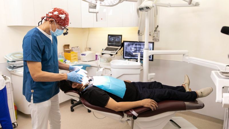
Yes, in selected cases. I can place the implant during the sinus lift when the remaining bone gives me reliable primary stability and I can protect the sinus membrane and graft. A small amount of residual bone does not automatically rule out simultaneous treatment, but it makes technique and case selection more demanding.
At the back of the upper jaw, the maxillary sinus may occupy the space where molar bone has reduced after tooth loss. A sinus lift raises the Schneiderian membrane and places graft material beneath it to create support for an implant. I treat that membrane as a wall that should remain intact; if it tears, it must be recognised and managed.
Published series show that simultaneous treatment has been performed in sites with limited residual height, but the evidence does not create an automatic threshold for every patient. Cha and colleagues reported 217 consecutive lateral sinus lifts with 462 simultaneous implants; 262 implants were placed where residual bone height was under 4 mm, and second-stage surgery occurred at about 6 months. Peleg, Garg and Mazor reported 2,132 implants placed with 731 sinus grafts; 20.4% of the implants were in sites with 1 to 2 mm of residual bone, with cumulative survival of 97.9% over follow-up extending to 9 years.
That evidence shows what experienced clinicians have achieved. It is not a tourism protocol and not a promise for your anatomy.
A multicentre trial in sites with 1 to 3 mm of residual bone reported 3 implant failures in the one-stage arm and 1 in the two-stage arm. The difference was not statistically significant, but the authors cautioned that one-stage treatment might carry a slightly higher failure risk at that height. I keep that caution in the decision.
Sinus-membrane perforation is the most common intraoperative complication. A European Workshop consensus reported a perforation rate of about 18%, while a related review reported a published range from 0% to 31.5%. When a perforation was recognised and repaired, a later meta-analysis reported implant survival of 97.1% beneath repaired membranes and 97.7% beneath intact membranes.
Those are published figures, not Westcoast figures. I will not invent a clinic-specific perforation or success rate.
4. Julia from Germany: A clinician-documented case
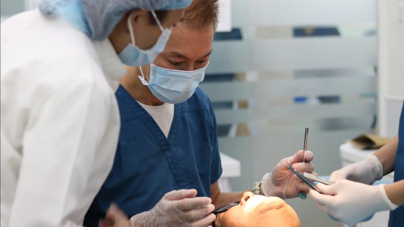
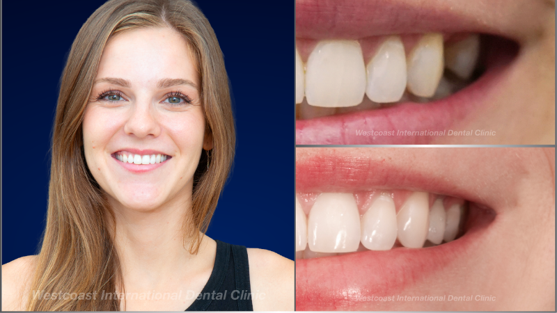
Julia from Germany presented with thin residual bone beneath the sinus after another dentist had declined the case. I planned and carried out the graft and implant during the same surgical visit because I could obtain stability in the remaining bone. Her treatment involved two clinic visits, with healing between them.
Those details come from the clinical case record and my account of the treatment. They are not words attributed to Julia’s video. One case cannot predict your result, healing or travel plan.
5. What type of bone graft might you need?
The right procedure depends on the shape of the defect. I do not choose a larger graft simply because it sounds more comprehensive. I match the technique to the missing bone and the final restoration.
5.1 Socket preservation
Socket preservation places graft material in an extraction socket to reduce ridge collapse after a tooth is removed. It can limit bone loss, but it cannot freeze the ridge at its original dimensions.
A systematic review measured mean horizontal ridge reduction of 3.79 mm during the first 6 months after extraction. A review of 22 randomised trials found that ridge preservation reduced horizontal collapse by 1.99 mm compared with unassisted healing. Other evidence confirms that resorption can be reduced but not completely stopped (MacBeth et al., 2017, Clinical Implant Dentistry and Related Research).
In a selected extraction site, I may place an implant with the graft. That remains a stability and case-selection decision.
5.2 Horizontal ridge augmentation
Horizontal augmentation rebuilds width when the ridge is too thin for an implant in the correct restorative position. The graft must remain stable and the soft tissue must close without tension.
A European Workshop consensus reported mean clinical width gain of 3.45 mm, with wound dehiscence and membrane or graft exposure ranging from 5% to 54% across the included literature (Jepsen et al., 2019, Journal of Clinical Periodontology). One study measured 3.01 mm of bone gain when sites remained closed and 0.56 mm when exposure occurred.
That is why I pay close attention to closure. A contained width defect may allow simultaneous implant placement; a larger defect may need staged reconstruction.
5.3 Sinus elevation
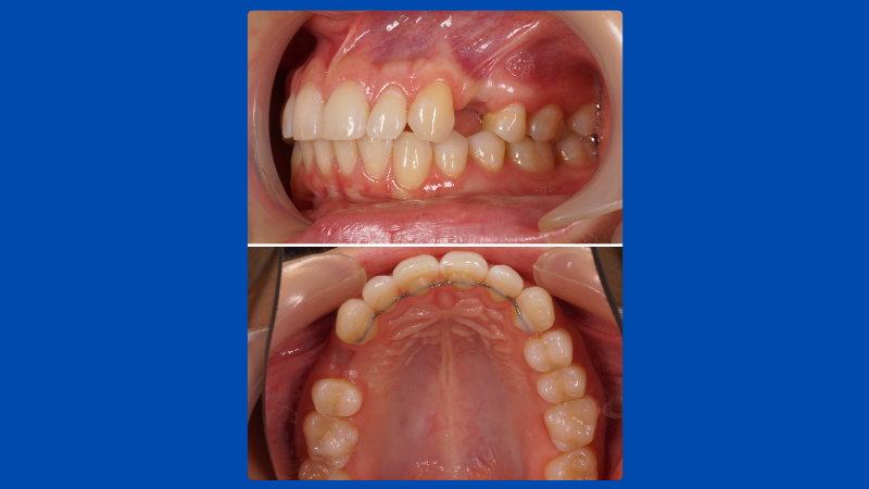
Sinus elevation is technically demanding because the membrane must be lifted and protected while the graft and, in selected cases, the implant are stabilised. I decide between a simultaneous and staged approach from the residual bone, implant stability, membrane condition and restorative position.
5.4 Vertical ridge augmentation
Vertical augmentation rebuilds height and is among the most technique-sensitive grafting procedures. I often stage tall vertical reconstructions because the site must create and protect a stable space beyond the existing bone envelope.
A systematic review reported a pooled mean vertical gain of 4.16 mm and an overall complication rate of 16.9%. In the same review, guided bone regeneration produced 4.18 mm of mean gain with 12.1% complications, block grafting produced 3.46 mm with 23.9% complications, and distraction osteogenesis produced 8.04 mm with 47.3% complications (Urban et al., 2019, Journal of Clinical Periodontology). These are pooled research results, not a height or outcome I can promise.
5.5 Complex revision
Revision begins with finding out why the previous dental implant failed. Infection, implant position, bite, hygiene, smoking, medical factors and a prosthesis that cannot be cleaned may all matter. Grafting again without understanding the first failure risks repeating it.
Replacement implant series show why I describe revision as less predictable. One study reported second-attempt survival of 88.84% from 390 of 439 implants and third-attempt survival of 74.19% from 23 of 31 implants (Zhou et al., 2016, International Journal of Oral and Maxillofacial Implants). Another series reported 71% survival for replacement implants (Grossmann and Levin, 2007, Journal of Periodontology).
I do not treat those figures as a forecast for an individual. They are a reason to diagnose the failure before planning another graft or implant.
6. Which bone graft material might be right for you?
I choose the material according to the defect, the volume needed, the need to maintain space and whether a donor site is justified. Autogenous bone, taken from you, brings living cells and remains an important reference material. It also requires a second surgical site and has limits in volume and donor-site morbidity.
A review described autologous bone as the current gold standard while also documenting those limitations (Haugen et al., 2019, Journal of Clinical Periodontology). Processed human-donor bone and well-documented bovine mineral can avoid a second harvest or add volume. Bovine mineral may help maintain space because it resorbs slowly, but a religious, ethical or personal objection is a valid reason to discuss another material.
I will tell you which material category I recommend and why. More graft is not automatically better.
7. What decides the difficult cases?
Primary closure, blood supply, space maintenance and graft stability decide whether regeneration has the conditions it needs. My job is to create and protect those conditions while controlling the risks that you bring to the site. As the defect becomes more complex, experience and execution matter more, not less.
Wang and Boyapati described these as the PASS principles: primary closure, angiogenesis, space creation or maintenance, and stability (Wang and Boyapati, 2006, Implant Dentistry). The framework supports predictable regeneration; it does not guarantee it.
In horizontal and vertical grafting, the work is experience and skill. Smoking and diabetes have to be controlled. Materials and technique matter, but as the case becomes harder, skill is what decides whether the biological conditions are protected. I treat the sinus membrane as a wall that should stay intact. The gum cannot open. If one of those conditions slips, you may get less bone – not a free pass.
– Dr Andrew H.F. Tsang
Smoking is associated with a higher risk of implant failure. A meta-analysis reported an odds ratio of 2.40 across 35,511 implants in smokers and 114,597 implants in non-smokers (Mustapha, Salame and Chrcanovic, 2022, Medicina). Diabetes is not an automatic exclusion, but control matters; another meta-analysis reported an implant-failure odds ratio of 1.78 for diabetes and 4.48 when comparing type 1 with type 2 diabetes (Al Ansari, Shahwan and Chrcanovic, 2022, Materials).
Tell me about smoking or vaping, diabetes control, immune conditions, anticoagulants, antiresorptive medicines and previous healing problems. Do not stop prescribed medicine because a webpage suggested it.
8. How will I decide whether you need a graft?
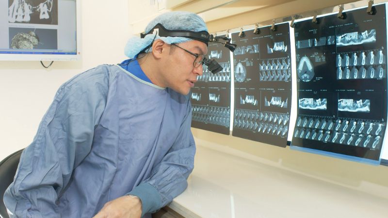
I first decide whether the site can hold an implant in the correct restorative position. I then decide whether grafting is needed and whether it can be combined safely with implant placement. Examination and imaging answer different parts of that question.
I assess the gums, adjacent teeth, bite, soft tissue and any signs of infection. CBCT imaging may show bone volume, the sinus and nearby nerves when it is clinically justified, but a scan does not make the decision by itself. Active infection may need treatment before grafting.
A remote review can narrow the options. The plan becomes final only after I examine you and review the imaging required for your case.
9. How long will healing take, and how many trips will you need?
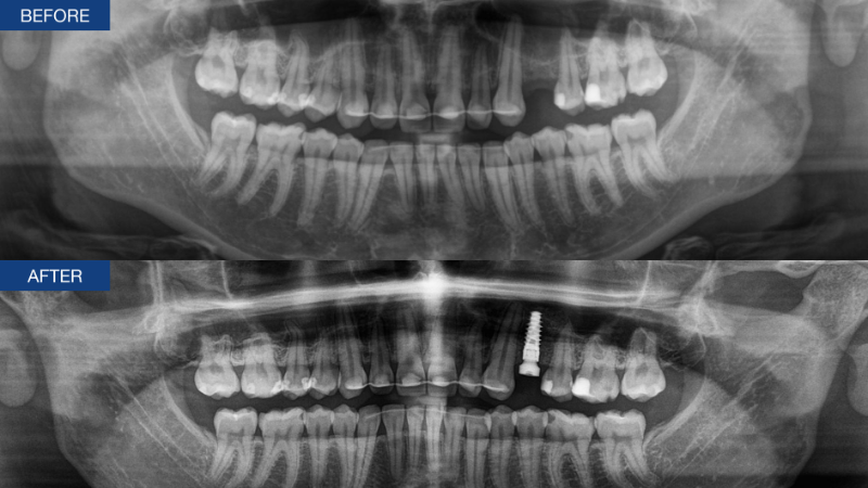
Your visit plan depends on whether I can combine the graft and implant, the size of the defect, early healing and when the implant is ready for restoration. In practical travel terms, simultaneous graft and implant placement will often mean two trips to Vietnam: one for grafting and implant placement, and another for the final restoration after healing. Staged treatment will often mean three trips: one for grafting, one for implant placement after the graft has healed, and one for the final restoration after implant integration. The exact schedule can change with early healing, temporary teeth and whether any restorative care is completed at home.
For larger lateral ridge defects, the clinical guideline cited above describes implant placement after 4 to 9 months of graft healing (Avila-Ortiz et al., 2023, Periodontology 2000). That is a research-based planning range, not a departure date or a promise for your case.
There is no universal safe-to-fly interval after every type of graft. I finalise travel advice after considering the operation, early healing, bleeding, swelling, temporary teeth and any sinus precautions. Feeling well is not the same as having a mature graft.
If you are travelling for treatment, leave enough time for the procedure, an early review and adjustment of any temporary tooth before onward travel. Do not assume the permanent tooth will be completed during a short surgical visit.
Read more: 3 Step Procedure for Having Dental Treatment in Vietnam
10. What temporary tooth can you have?
A temporary tooth is not the final crown. It protects appearance and limited function while the graft and implant are left to heal. I choose it according to how well pressure can be kept away from the surgical site.
A removable temporary may be the safest option when a fresh graft must not be loaded. A fixed provisional restoration is possible in selected cases, but I do not assume it before assessing stability and bite. If you clench or grind, tell me before treatment.
11. What are the risks and warning signs?
Every graft can develop infection, exposure, insufficient bone formation or delayed healing. An implant placed at the same time can also fail to integrate. If that happens, I assess the cause before deciding whether removal, more healing, revision or another restorative plan is appropriate.
Contact the clinic and seek local dental or medical care if you develop:
- Pain that increases after it had started to settle, spreading swelling, fever, pus or a foul taste;
- An opening in the gum, visible membrane or graft, or material being lost;
- Bleeding that continues despite the instructions you were given;
- Numbness, tingling or another change in sensation that persists or worsens;
- After an upper-jaw or sinus procedure, new sinus pain, pressure, nasal symptoms or fluid passing between the mouth and nose;
- An implant that feels mobile or a temporary tooth that is pressing on a site meant to remain unloaded.
Small granules can occasionally appear after a particulate graft. Do not poke the site. An open wound, ongoing material loss or worsening symptoms needs examination.
Antibiotics are not automatic. Prescribing depends on the procedure and your clinical situation.
12. What happens after treatment?
Follow the cleaning, diet, medicine and activity instructions given for your procedure. Do not smoke or vape, chew on the graft, let a temporary tooth press on it, or stop prescribed medicine without speaking to the relevant clinician. Attend the planned reviews even if you feel well.
Before you leave Vietnam, the clinic can prepare a treatment summary, relevant images, graft and implant details where applicable, aftercare instructions, emergency contacts and the next review plan. If a dentist at home may complete the restorative stage, confirm that they are willing to do so before you depend on that arrangement. I cannot promise that a clinician I have not met will accept the case.
13. What should you send before an enquiry?
Send a recent panoramic X-ray or CBCT if you have one, clear photographs of your teeth and bite, a short dental and medical history, your medicines and allergies, smoking status, and records of previous implants, grafts or failed treatment.
Ask for a written plan that explains:
- Whether the graft and implant may be combined or should be staged, and why;
- What remains preliminary until you are examined;
- The proposed procedure and graft-material category;
- The likely healing sequence and number of visits;
- The temporary-tooth plan and how pressure will stay off the graft;
- Travel and sinus precautions;
- What happens if the graft or implant does not heal as planned;
- Which records your dentist at home will receive.
A remote review supports planning. It is not a final diagnosis, a confirmed timeline or a promise that grafting or implant placement will be possible.
14. Frequently asked questions
14.1 Can you have a bone graft and an implant at the same time?
Yes, when the remaining bone can stabilise the implant and the defect can be protected. I favour simultaneous treatment when those conditions are present. I stage the graft when they are not.
14.2 Can you have a sinus lift and an implant in the same visit?
Yes, in selected cases where I can achieve primary stability and manage the sinus membrane safely. Limited residual bone makes the procedure more demanding; it does not create an automatic yes or no.
14.3 Can you have dental implants without a bone graft?
Yes, if there is adequate bone in the correct position for the final restoration. Examination and appropriate imaging decide that. A graft is not automatic.
14.4 Can you fly after a dental bone graft?
There is no single interval that applies to every graft. I give travel advice after considering your procedure, early healing, swelling, bleeding, temporary teeth and any sinus precautions.
14.5 Can the final crown be completed at home?
It may be possible if your dentist at home agrees and has the necessary records and compatible components. Confirm that arrangement before treatment. The clinic can also plan the restorative visit in Vietnam.
14.6 What if the graft does not heal or the implant does not integrate?
I investigate the cause first. The next step may involve more healing time, infection management, graft revision, implant removal, a modified implant plan or a non-implant restoration. Revision may require more visits and is generally less predictable than first treatment.
Send an enquiry
Send your images and history through westcoastinternational.com. I would rather tell you that a graft is unnecessary than add one to your plan. If you do need grafting, I want the procedure that matches the defect and the final tooth.
– Dr Andrew H.F. Tsang
Bone grafting and implant placement are decided case by case. Simultaneous treatment is suitable when the remaining bone can stabilise the implant and the graft can be protected. Staged treatment is chosen when those conditions are not met. A remote X-ray starts the discussion. The final plan is made only after examination and proper imaging.
Sources cited in this draft
- Al Ansari, Shahwan and Chrcanovic. Materials. 2022.
- Avila-Ortiz, Chambrone and Vignoletti. Journal of Clinical Periodontology. 2019.
- Avila-Ortiz, Couso-Queiruga, Stuhr and Chambrone. Periodontology 2000. 2023.
- Cha, Kim, Nowzari, Chang and Ahn. Clinical Implant Dentistry and Related Research. 2014.
- Díaz-Olivares and colleagues. Journal of Clinical Medicine. 2024.
- Felice, Pistilli, Piattelli, Soardi, Barausse and Esposito. European Journal of Oral Implantology. 2014.
- Grossmann and Levin. Journal of Periodontology. 2007.
- Haugen, Lyngstadaas, Rossi and Perale. Journal of Clinical Periodontology. 2019.
- Jepsen, Schwarz, Cordaro, Urban, Simion, Rocchietta and colleagues. Journal of Clinical Periodontology. 2019.
- MacBeth, Trullenque-Eriksson, Donos and Mardas. Clinical Implant Dentistry and Related Research. 2017.
- Machtei. Journal of Periodontology. 2001.
- Mustapha, Salame and Chrcanovic. Medicina. 2022.
- Peleg, Garg and Mazor. International Journal of Oral and Maxillofacial Implants. 2006.
- Raghoebar and colleagues. Journal of Clinical Periodontology. 2019.
- Sanz-Sánchez, Ortiz-Vigón, Sanz-Martín, Figuero and Sanz. Journal of Dental Research. 2015.
- Tan, Wong, Wong and Lang. Clinical Oral Implants Research. 2012.
- Urban, Montero, Monje and Sanz-Sánchez. Journal of Clinical Periodontology. 2019.
- Wang and Boyapati. Implant Dentistry. 2006.
- Zhou and colleagues. International Journal of Oral and Maxillofacial Implants. 2016.

Book An Appointment Now!
Connect With Us!
Healthcare Group Members


Copyright All Rights Reserved